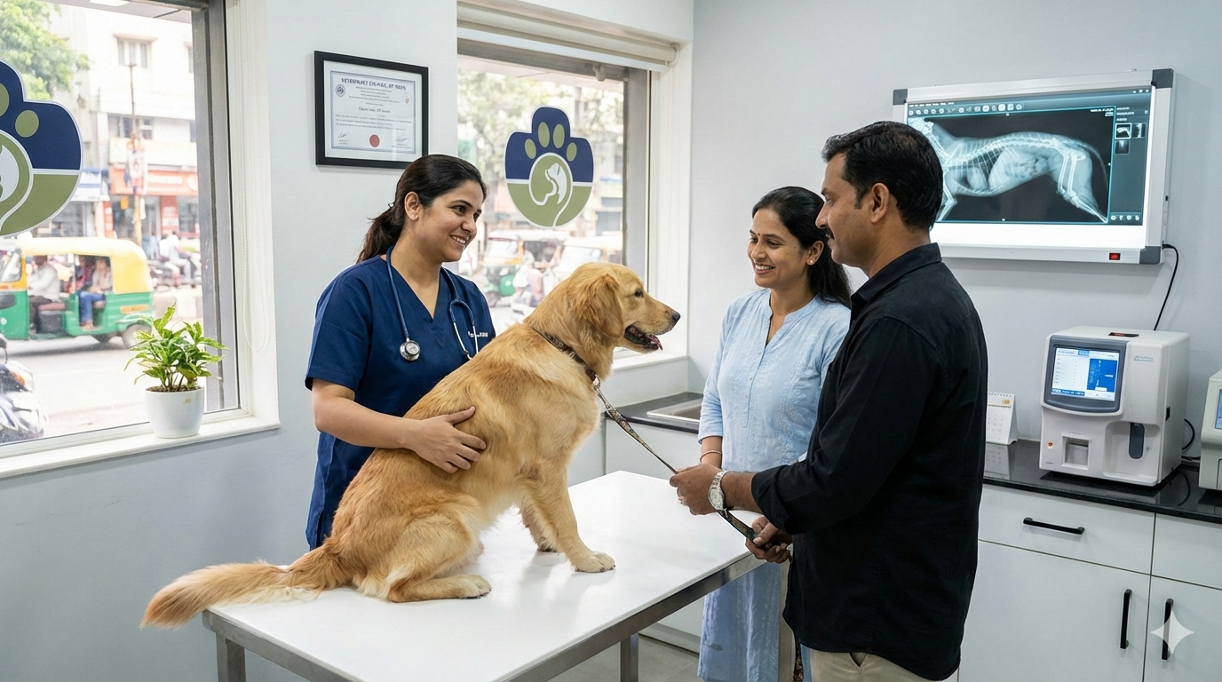
The Veterinary Clinical Complex (VCC) of IIVER is catering the need of farmers and providing round the clock service to the farmers for treatment of their animals. Different types of clinical cases brought are Cattle, Buffalo, Sheep, Goat, Pig and Poultry etc. The cases after registration are examined by the experts using different scientific methods such as laboratory tests and diagnostic imaging techniques (X-ray and ultrasound) and correct line of treatment is adopted to manage the clinical cases.
Services Offered
- 24/7 Veterinary Care: We understand that emergencies can arise at any time. Our VCC operates around the clock to provide prompt veterinary attention to the animals of farmers in our community.
- Extensive Species Coverage: Our services cater to a wide variety of animals, including cattle, buffalo, sheep, goats, pigs, and poultry.
- Advanced Diagnostics: We utilize cutting-edge scientific methods to ensure accurate diagnoses. This includes advanced laboratory testing and diagnostic imaging techniques such as X-ray and ultrasound.
- Specialized Care: The VCC is comprised of dedicated sections focusing on Surgery, Medicine, Gynecology, and Small Animal care. Each section is staffed with specialists who possess in-depth knowledge and expertise in their respective fields.
- Farmer Support: In addition to treating animals, we offer valuable advisory services to farmers. This guidance empowers them to implement proper animal care practices, ultimately contributing to the overall health and productivity of their livestock.
MEDICINE SECTION
Our team effectively diagnoses and treats various medical conditions in animals. Common ailments addressed include Mastitis in cattle, Canine Parvoviral disease in puppies, and Carbohydrate Engorgement in cattle
MASTITIS
Bovine mastitis, an inflammation of the mammary gland, is the most common disease of dairy cattle causing economic losses due to reduced yield and poor quality of milk.
Clinical Signs : Inflammation of udder and systemic sign fever, Decreased Milk yield , Swelling in udder , milk flakes ,Change in milk colour , Fibrosis in chronic case.
Diagnosis: History, clinical signs, California Mastitis Test (CMT), strip cup test, Cultural Sensitivity Test (CST).
Treatment: Anti-histamine Broad spectrum antibiotics, anti-inflammatory, supportive therapy etc.

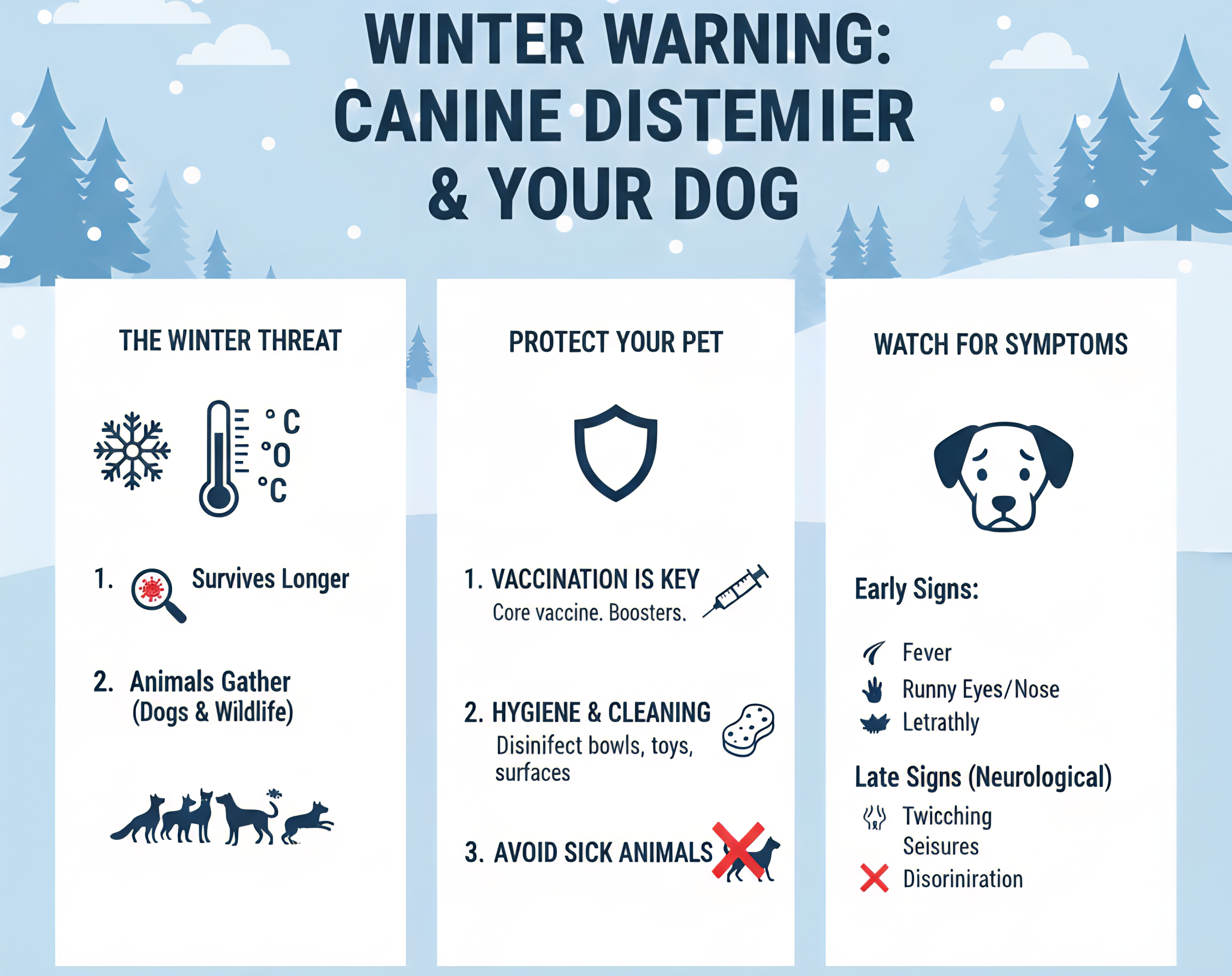
CANINE PARVOVIRAL DISEASES
Canine Parvo in puppies is caused by the canine parvovirus. This virus is highly contagious and spreads through direct contact with an infected dog or by indirect contact with a contaminated object. Puppy is exposed to the parvovirus every time he sniffs, licks, or consumes infected feces. Indirect transmission occurs when a person who has recently been exposed to an infected dog touches your puppy, or when a puppy encounters a contaminated object, like a food or water bowl, collars and leashes.
Clinical signs: Anorexia, fever, vomition, foul smell bloody diarrhoea, fever, unvaccinated dogs of less than 3 month are more susceptible.
Treatment: Symptomatic treatment, Broad spectrum antibiotics, antacid, anti-emetic drugs. Supportive therapy like vitamin-B complex and Ascorbic acid.
Prevention: Vaccination at the age of 6-8 week. Booster dose after 21 days and then annually vaccination.
CARBOHYDRATE ENGORGEMNT
Grain overload is most common in cattle that accidentally gains access to large quantities of readily digestible carbohydrates, particularly grains. Grain overload also is common in feedlot cattle when they get approached accidentally to heavy grain diets too quickly. Wheat, barley, and corn are the most readily digestible grains; oats are less digestible.
Clinical sign: Inapptance to anorexia, decreased ruminal motility, bloat, constipation followed by diarrhoea, decrease in rumen pH. There can be difficulty in respiration with increase in respiration rate up to 60–90 breaths/min. Heart rate usually get increased in accordance with severity of the academia. Prognosis is poor for cattle showing sign of tachycardia.
Treatment: In severe cases rumenotomy or rumen lavage is indicated combined with fluid therapy. In moderate cases, alkalizing agents (soda bicarbonate) is advised.

SURGERY SECTION
Our skilled surgeons manage a range of surgical procedures, including those for Foreign Body Syndrome in ruminants, Ear Hematoma in dogs, Obstructive Urolithiasis in male calves, Umbilical Hernias in calves, and Teat Fistula in dairy animals.
FOREIGN BODY SYNDROME
The incidence of ingestion of foreign bodies along with food is very common in ruminants. The common ingested foreign bodies are nails, wires, leather, ropes etc. It may cause perforation of reticulum thereby causing traumatic reticulo-peritonitis. The most common clinical findings include decreased feed intake, tympany, decreased rumen motility, mild fever, scanty black feces, and signs of pain. Sharp objects may cause peritonitis and abdominal adhesion formation. Reduced milk production, symptoms of stomach discomfort, and rumenoreticular atony are examples of complications of ingested metallic objects. Radiography (X ray) is commonly used to diagnose metallic foreign bodies in the reticulum. Usually surgical procedures to open the rumen i.e., rumenotomy is performed to takes the foreign bodies out.
Sometimes, these foreign bodies may cause rupture of the diaphragm and causes diaphragmatic hernia. A diaphragmatic hernia is characterized by the protrusion of abdominal viscera into the thoracic cavity through diaphragm. It primarily may happen as a result of increased intra-abdominal pressure caused by advanced stage of pregnancy or at the time of parturition. It could also happen as a result of trauma caused by metallic object. The repair of ruptured diaphragm is performed into two stages. In first stage, rumenotomy is carried out to evacuate the rumen and in second stage, repair of diaphragm is performed.
EAR HEMATOMA
Collection of blood in a dog's ear pinna is called an ear hematoma.
Causes: An ear infection or other ear irritation, such as allergies or ticks infestation, is the most frequent cause of an ear hematoma in dogs. Additional reasons include bite wounds, yeast infections, immunological disorders, blood-clotting disorders, skin conditions, pinna trauma, ear mites, etc.
Clinical signs: The pinna will become thick and spongy and the accumulated blood may be fresh or clotted. One part of the ear or the whole ear may become swollen. Because they are so irritable to dog, ear hematomas need to be treated right away. The hematoma may harm the ear tissues around it if treatment is not received, leading to an ear shaped like a cauliflower that may clog the ear canal.
Treatment: The most effective method for chronic or recurring hematomas is surgical drainage. Drain tubes or sutures along with opening the ear can be used to manage surgery. Early intervention will reduce the chance of scarring and avoid pressure soreness and a large ear flap.
OBSTRUCTIVE UROLITHIASIS IN MALE CALF
In feedlots, it ranks as the fifth most frequent cause of mortality. Male calves are more likely to be affected than female ones, and it is more likely to happens during the winter.
Causes: Obstructive urolithiasis is caused by calculi that obstruct the passage of urine in the urinary system.
Clinical signs: In cases where the bladder is intact, the common clinical signs are anuria, inappetence, dull and anxious look, kicking at the ventral abdomen, and persistent attempts at micturition, etc. In cases where the bladder is ruptured, the signs included bilateral distension of the abdomen with fluid thrill, sunken eyes, and no attempt at micturation. Rectal prolapse may also be present in some cases. Animal may die if not treated properly and on time.
Treatment: For the surgical treatment of obstructive urolithiasis, tubecystostomy is proved to be an effective treatment. In this, a Foley’s catheter is inserted into the urinary bladder through surgical opening on ventral abdomen region. This catheter facilitates the flow of urine formed in urinary bladder to outside the body. Urethral calculi in urinary bladder are removed by surgical procedure and those present in urethra are treated by providing ammonium chloride orally.
UMBILICAL HERNIAS
In calves, umbilical hernias are the most prevalent birth abnormality.
Causes: Congenital
Clinical signs: With the exception of reducible umbilical hernias, which allow the contents of the hernia to be readily pushed back into the belly, calves with uncomplicated hernias may appear quite normal. Hernia infections in calves can cause illness and manifest as fever, inappetence, and slow development. Calves get physical examinations in an effort to lessen the amount of hernia. An infected stalk inside the hernia sac or around the umbilicus may occasionally be palpable.
Diagnosis: To diagnose hernia ring along with presence of infection can be confirmed by ultrasonography.
Treatment: To properly feel these structures, the calf may need to be sedated and placed on their dorsal recumbancy. The surgical method used to repair the hernia ring is overlapping technique using non-absorbable suture material. Daily antiseptic dressing along with antibiotic, analgesic and abdominal bandage is advised till removal of skin suture.
TEAT FISTULA IN DAIRY ANIMALS
Teat lacerations were a common occurrence in dairy cattle reared in zero grazing systems and caused losses in milk production (Nichols, 2008). Laceration if deeper then even teat canal gets opened and milk will start flowing through the affected portion leading to teat fistula. Deeper lacerations involving the teat canal required prompt suturing (within 6 hours of trauma) of the defects (Roberts and Fishwick,2010). Local anesthesia ring block techniques facilitated surgical repair of lacerated and traumatized tissues of the udder.
Causes: Most common causes of teat lacerations are direct trauma by sharp wire or self foot, particular in pendulous teat or injury during suckling by calf.
Clinical Signs: Deep laceration causes formation of teat fistula which is an abnormal opening on the teat surface from which continue flow of milk occurs direct from main teat cistern. Teat fistula cases should be managed as early as possible to prevent complications like mastitis, sepsis or necrosis of teat.
Treatment: First of all, animal is sedated and restrained, affected teat is washed with antiseptic solution and suturing is done properly. A prosthetic tube is placed properly into the teat which aid in removal of milk from udder, healing of fistulous tract and prevent the blockage of teat canal during healing.

GYNAECOLOGY SECTION
Our team offers comprehensive reproductive health services for female animals. Conditions addressed include Anoestrus, Repeat Breeding, Retention of Placenta, Dystocia, Metritis, Uterine Torsion, Incomplete Cervical Dilation (ICD), Transmissible Venereal Tumor (TVT), Uterine Inertia, Prolapse, Pyometra, Hydroallantois, and Hydroamnion.
ANOESTRUS
Absence of overt signs of estrus
Causes: Congenital abnormalities; pathological conditions of uterus like pyometra, metritis; luteal cyst; malnutrition; heat stress; silent heat
Clinical signs: Animal does not show any symptoms of heat.
REPEAT BREEDING
A normal cyclic female with apparently normal genitalia mated in 3 or more consecutive estruses with fertile bull or inseminated artificially with fertile bull semen if fails to conceive is called repeat breeder.
Causes: Delayed ovulation, anovulation, fertilization failure, luteal insufficiency, endometritis, ovario-bursal adhesions, follicular cyst, improper heat detection, environmental stress, improper timing of AI, nutritional deficiency.
Clinical signs: animal comes in heat at regular intervalsbut not conceive.
RETENTION OF PLACENTA
Non separation and failure of expulsion of fetal membrane within a certain time limit (cow:8-12 hrs)
Causes: hypocalemia, deficiency of oxytocin
Clinical signs: retained placenta hangs from vagina
DYSTOCIA
Difficulty in parturition
Causes: both maternal (small pelvis, uterine inertia) and fetal (fetal oversize, abnormal presentation, position and posture)
Clinical signs: animal continues to straining but second stage of labor not commences.
METRITIS
Inflammation of uterus involving all the three layers
Causes: occurs after abortion or retention of placenta; poor hygienic conditions at time of parturition; unsterilised instruments used for AI
Clinical signs: pus or mucopurulent discharge from vagina, systemic signs like decrease in appetite and milk yield; fever
UTERINE TORSION
Rotation of uterus along its longitudinal axis
Causes: sudden movement of dam or fetus, weakness of ligaments
Clinical signs: animal continue to straining but no progress in parturition process, frequent sitting and standing of animal
INCOMPLETE CERVICAL DILATION (ICD)
When lumen of cervical canal not dilate completely
Causes: hormonal imbalance, hard cervix
Clinical signs: animal continue to strain but no progress in parturition process
TRANSMISSIBLE VENEREAL TUMOR (TVT)
Also known as venereal granuloma is a benign reticuloendothelial tumor of dog that mainly affects the external genitalia and occasionally the internal genitalia.
Causes: transmit via coitus, real cause unknown possibly of viral etiology
Clinical signs: friable mutilobulated cauliflower like growth on penis in males and anterior vagina in females, bleeding
UTERINE INERTIA
Lack of normal physiological contractions of uterine muscles during and after parturition
Causes: Hypocalcemia; Small litter (insufficient oxytocin); Exhaustion of uterine muscles; Lack of exercise
Clinical signs: Relaxation of soft structures of pelvis, Discharge of mucus from vulva, History of prolonged dystocia
PROLAPSE
Protrusion of the whole or part of organ through the vulva/ natural opening is called prolapse (also k/a Phool dikhana)
Causes: Increase intra-abdominal pressure; Deficiency of Ca and disturbed Ca:P ratio; Urinary infection, vaginal injuries, Excessive traction, RFM etc
Clinical signs: prolapsed mass hang out from vagina
PYOMETRA
Accumulation of purulent material within the uterine lumen of intact bitches.
Causes: hormonal imbalance; bacteria like Escherichia coli, Staphylococcus, Pseudomonas, Proteus spp.
Clinical signs: foul smelling sanguineous to mucopurulent vaginal discharge, lethargy, depression, inappetence/anorexia, polyuria, polydipsia, vomiting and diarrhoea.
HYDROALLANTOIS AND HYDROAMNION
A pathological condition of the pregnant animal characterized by excessive accumulation of fluid within the amniotic or allantoic cavity.
Causes: Cystic, hydronephrosis or dysfunction of fetal kidneys, Vit. A deficiency
Clinical signs: Distended uterus and enlarged abdomen, Anorexia, lack of rumination, constipation; Elevated pulse rate, restlessness, expiratory grunt, stiff gait; Animal lies on sternum (bloated bull frog appearance)
Conclusion
The IIVER Veterinary Clinical Complex is a well-equipped and well-staffed facility that stands firmly behind its commitment to animal health. We offer advanced veterinary services for a wide range of animals, catering to both farmers and pet owners. If you have any concerns regarding your animal's health, please do not hesitate to contact us. We are here to serve you and your animal companions.


